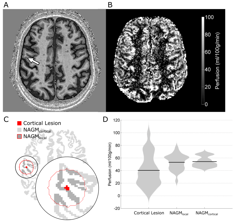
 In our latest paper in European Radiology, we use ultra-high field MRI to explore the clinical usefulness of an emerging technique for measuring perfusion that doesn’t involve the injection of contrast agents. Instead, in Arterial Spin Labeling (ASL) scans, we magnetically label the blood and then image it as it flows into the tissue, allowing us to quantify perfusion. We developed this technique for a specific clinical application: to better understand the role of small cortical lesions in Multiple Sclerosis (MS).
In this technical development report, we present an optimized ultra-high-field ASL MRI acquisition that achieves high spatial resolution and low signal distortion. We assess the feasibility of using this imaging strategy to measure perfusion in MS cortical lesions, and demonstrate that our approach is sensitive to focal hypoperfusion in these small lesions. Because ASL MRI is safe and non-invasive, this type of imaging approach may facilitate the longitudinal study of acute lesion formation and development with frequent repeated scanning, allow us to test new therapeutic strategies, and give us better understanding of the heterogenous disease course in MS.
This excellent work was primarily accomplished by Rich Dury and Yasser Falah as part of their PhD research. For more details, the manuscript is available (open access) here: https://rdcu.be/8n4G