Two new publications in collaboration with SPiN lab

Announcing the availability of two new publications in collaboration with the SPiN lab, lead by Cesar Caballero-Gaudes at the Basque Center on Cognition, Brain and Language in Spain! Both focus on the preprocessing and analysis of breath-hold fMRI data to measure cerebrovascular reactivity (CVR) and hemodynamic lag:

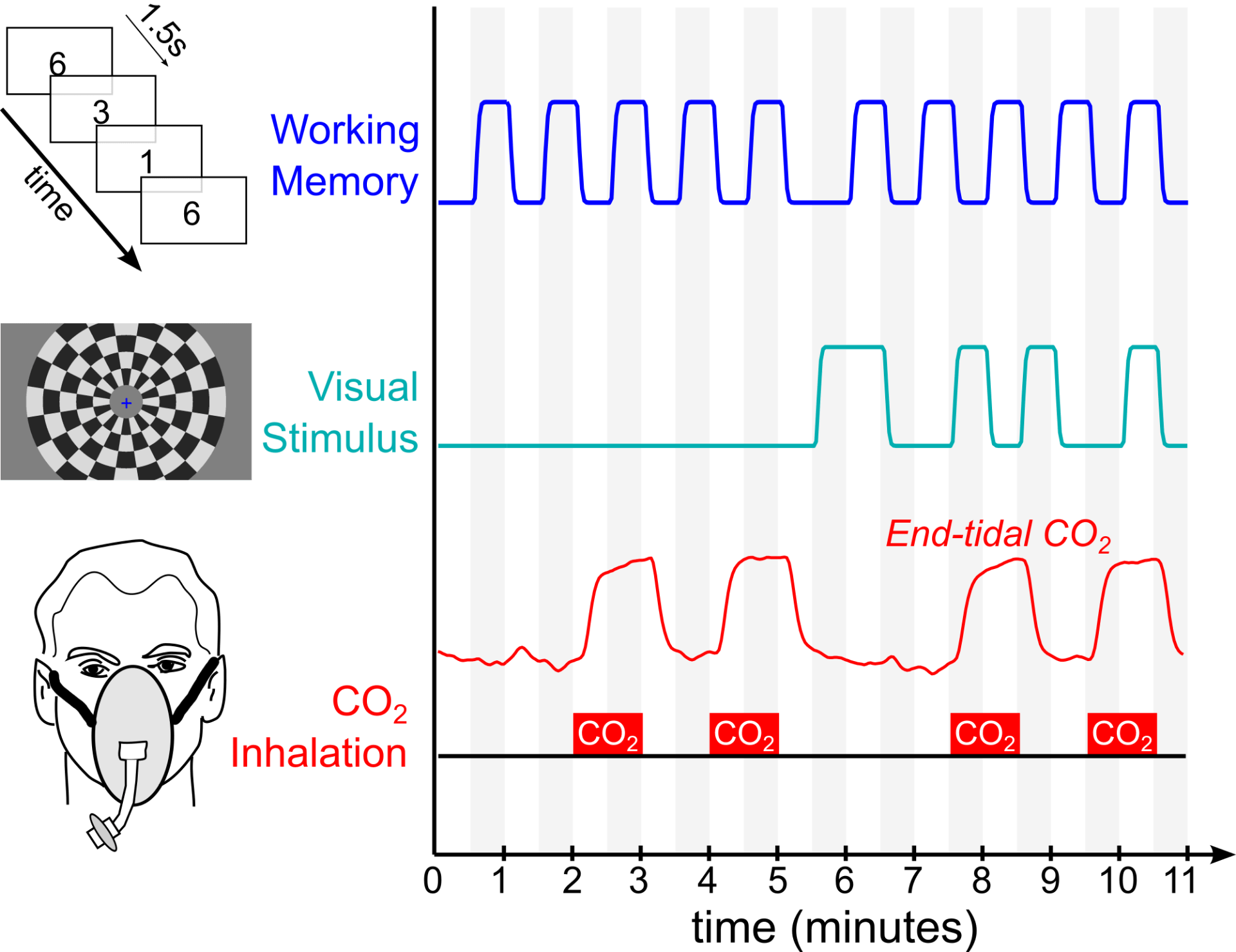
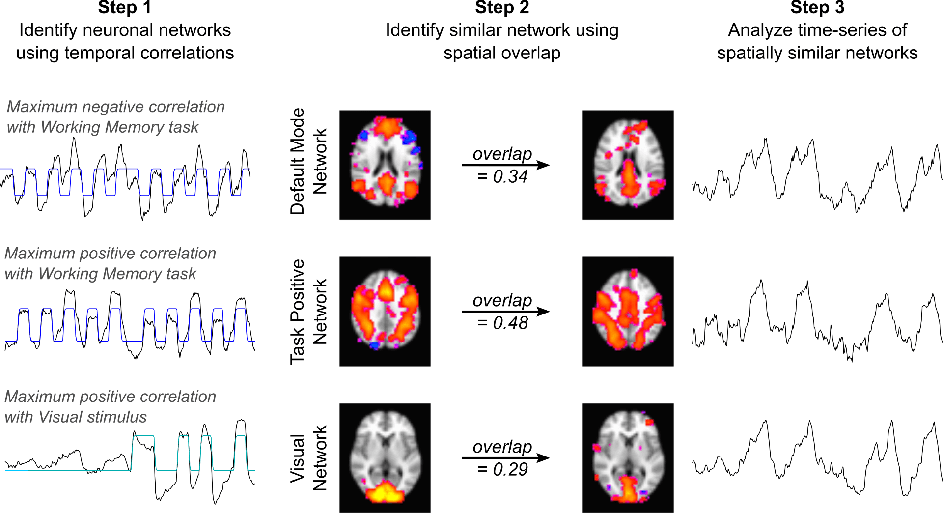
BCBL PhD student Stefano Moia and ANVIL postdoc Rachael Stickland co-authored an IEEE paper describing our lagged Generalized Linear Model approach for estimating the temporal delay between systemic carbon dioxide changes and the local BOLD fMRI response, mapping CVR and lag at the voxel level. The approach simultaneously fits other fMRI confounds, such as head motion, to better isolate the physiologic effects of interest. Download the pdf here!

Using the BCBL EuskalIBUR dataset, which uniquely studies breath-hold CVR across 10 repeated weekly MRI sessions, a new preprint in BioRxiv demonstrates the benefits of multi-echo data and compares multi-echo ICA denoising strategies for reliably mapping CVR and lag. Download the pdf here!

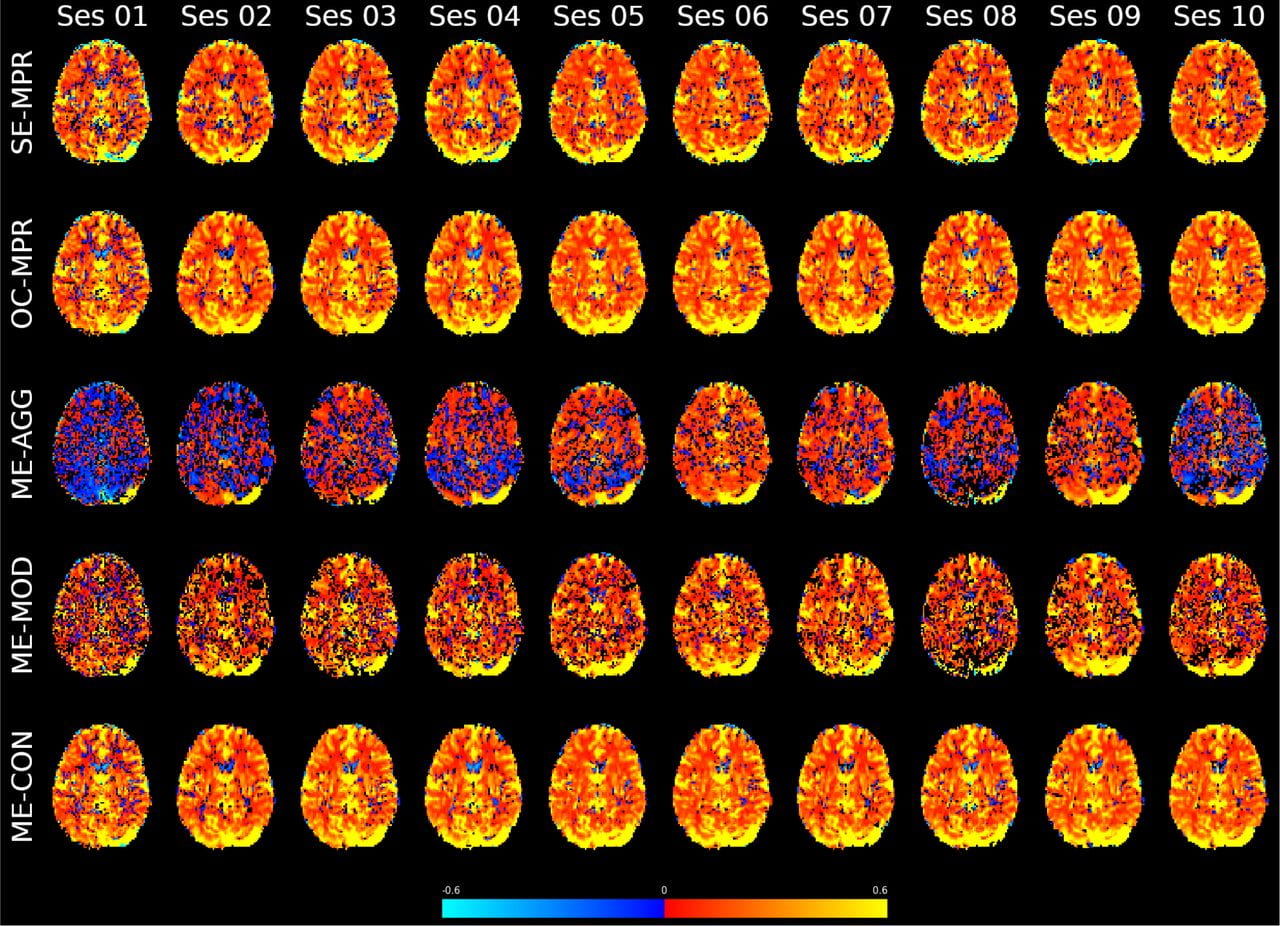
CVR maps for an example subject, scanned weekly for 10 weeks. Each row reflects a different use of multi-echo fMRI data, either combining information across the echo times or using the relationship across echo times and Independent Component Analysis (ICA) to differentiate the CVR effects from highly collinear noise confounds.

Research featured at ISMRM Annual Meeting

Recent work from the lab, lead by first author Dr. Rachael Stickland, was featured in the closing highlights session of the annual meeting of the International Society for Magnetic Resonance in Medicine. The meeting, held virtually for the first time, brought together over 5 thousand clinicians and scientists in the field of MRI and medical imaging for a week of talks, poster presentations, and virtual discussion. Dr. Stickland’s work was selected and discussed by Dr. Kevin Murphy (Cardiff University Brain Research Imaging Center), and demonstrates our lab’s proposed technique for characterizing the responsiveness of brain blood vessels using a simple, feasible, breathing task protocol during scanning. Congrats to all the authors!

Congratulations to Apoorva Ayyagari on completing her Masters Degree!

The Bright Lab celebrates Apoorva Ayyagari’s completion of her MS degree in Biomedical Engineering at Northwestern! After 2 excellent years as a core member of our lab, Apoorva successfully defended her MS thesis, titled “Understanding noise in spinal cord BOLD fMRI data with a breath-hold paradigm to investigate feasibility of studying vascular reactivity“. In this work, she rigorously assesses cardiac and respiratory noise, motion confounds, and co-linearity between different physiologic signals in spinal cord imaging data. Spinal cord fMRI data are notoriously challenging to work with due to all of these artifactual signals, and this thesis reflects excellent progress in understanding and modeling these factors. Apoorva’s many contributions in the lab will continue to benefit our ongoing projects in mapping cerebrovascular reactivity amplitude and latency, standardizing how physiologic data is collected during MRI scanning, preprocessing spinal cord fMRI data, and quantifying relationships between different sources of signal variance in these data. Although we are extremely sad to see her go, we wish Apoorva the very best as she starts her new job in human factors engineering in Evanston. Congrats Apoorva!

Demonstrating network-level interactions between neural and vascular function

Our most recent work demonstrating that the brain’s vasculature is regulated to mimic neural networks is now published in Neuroimage: https://doi.org/10.1016/j.neuroimage.2020.116907. This work was performed in collaboration with Prof. Kevin Murphy’s lab at Cardiff University Brain Research Imaging Center in the UK.

Using fMRI, we simultaneously administer two neural paradigms (a working memory task and a visual stimulus) and one “vascular” paradigm that dilates blood vessels systemically (inhaled CO2). We then averaged together 30 fMRI datasets and used Independent Component Analysis to decompose the average dataset into network components. We readily identify three “neural networks” that show strong temporal correlation with the neural stimulus paradigms. These represent the Default Mode Network, Task Positive Network, and Visual Network – three robust and commonly observed functional brain networks expected to be activated or deactivated by our neural paradigms. However, we also see three additional components with similar network structure, and these three networks predominantly reflect the vascular stimulus design.

Our results demonstrate, for the first time, pairs of spatially similar neural and vascular brain networks. This suggests that the brain’s vasculature may be regulated to support specific brain networks, which must be taken into account to interpret fMRI studies of functional connectivity.

Dr. Rachael Stickland receives NUCATS pilot grant

Congratulations to postdoctoral fellow Dr. Rachael Stickland on her success in the NUCATS Pilot Grant program! Over the coming year, Dr. Stickland will be assessing a multi-parametric imaging protocol focused on cerebrovascular function, testing reliability in healthy participants then translating into patients. This project will use the RespirActâ„¢ to deliver carbon dioxide in a carefully controlled, breath-by-breath basis during concurrent MRI scanning to characterize the associated vasodilatory response. More information about NUCATS can be found on their website.

Funding from the Craig H. Neilsen Foundation to study neural plasticity in Spinal Cord Injury

The Craig H. Neilsen Foundation has awarded Dr. Molly Bright a SCIRTS Pilot Grant to use imaging to characterize neurovascular plasticity in spinal cord injury. Working with Dr. Milap Sandhu, PT PhD at the Shirley Ryan Ability Lab, we will employ fMRI and cerebrovascular imaging techniques to characterize how one session of Acute Intermittent Hypoxia impacts brain and spinal cord physiology. Acute Intermittent Hypoxia is an emerging intervention that transiently improves motor function in individuals with spinal cord injury; although large clinical trials are getting underway, we aim to better understand the mechanisms for these functional improvements in order to optimize or tailor the intervention. This study will span two years, and recruitment is soon beginning for individuals with incomplete cervical spinal cord injury as well as uninjured control participants. Mark Hoggarth, DPT (left) will be joining the lab later this fall to take the lead on spinal cord imaging and analyses. Welcome Mark, and stay tuned for details on how to get involved with the study!

Open Position for a Postdoctoral Fellow in SCI

Update: An offer has been extended and accepted. Please see the Openings page for details on current positions.

We are excited to announce an opening for a full-time Postdoctoral Fellow to characterize neurovascular plasticity in Spinal Cord Injury. This study uses MRI of the brain, brainstem and spinal cord to probe the physiological mechanisms underpinning a new therapeutic intervention for SCI, which is currently undergoing clinical trials at the Shirley Ryan AbilityLab. This position is for 2 years with the possibility to extend.

For further details, please see the full description on the Openings page. Interested applicants are encouraged to get in touch with Prof. Bright with their questions prior to submitting an application. It may also be possible to arrange for an informal discussion in person during the ISMRM annual meeting in Montreal. (Applications received prior to June 17, 2018 will receive full consideration.)

We look forward to hearing from you!

Molly Bright elected to the ISMRM Brain Function Study Group committee

PI Molly Bright has just been elected as secretary of the Current Issues in Brain Function Study Group of the International Society for Magnetic Resonance in Medicine. She will be introduced formally at the annual meeting in Montreal, and will have a 4-year tenure advancing through the committee positions until finishing as Chair in 2023.

All interested in imaging brain function are invited to attend the study group meeting in Montreal, which will be held on Tuesday, May 14th, 13:30-14:30. We’re looking forward to working closely with the committee and members of the study group!


Welcome Jasmine Vu, Apoorva Ayyagari to the lab!

ANVIL welcomes two new students in Biomedical Engineering:

Jasmine Vu is an incoming PhD student, having completed her Bachelors degree in Bioengineering at the University of Washington with a concentration in Diagnostics and Therapeutic Instruments. She was an active member of Bioengineers Without Borders, sparking her interest in clinical translation of new technologies, and has extensive research experience in optical coherence tomography through her work in the UW Biophotonics and Imaging Lab under Dr. Ruikang Wang. Jasmine will be co-supervised by Prof. Bright and Prof. Rad, developing fMRI as a tool for studying brain function after deep brain stimulation (DBS) implantation. Her research will radically enhance our understanding and optimization of DBS for treating Parkinsons Disease.

Apoorva Ayyagari is a new Masters student in the lab, joining us from Oregon State University where she recently completed her Bachelors degree in Bioengineering. She has contributed to research in the Social Mobility Lab there, analyzing the movement patterns of young children with and without developmental disabilities. Here at Northwestern, Apoorva will be using MRI techniques to study oxygen dynamics in the human spinal cord, and how this is affected in spinal cord injury.

We are excited to have these two exceptional individuals join the team!