Multiple ANVIL successes to celebrate!

Congratulations are in order for three outstanding accomplishments from ANVIL lab members this past month!

Becca Clements (2nd year PhD student in BME) was awarded a 3-year fellowship from the NSF Graduate Research Fellowship Program! This will fund her exploration into the organization of vascular regulation in the human brain, and particularly the presence of long-distance “vascular networks” that may support typical functional neural networks.

Kim Hemmerling (4th year PhD student in BME) received a stellar 6%ile score on her NIH F31 Predoctoral Fellowship application! This score reflects the amazing potential for the spinal cord fMRI methods she has been developing in our lab, and specifically towards looking at atypical upper limb movement patterns post-stroke.

And finally, Max Wang (Postbac Research Technologist) has been awarded a prestigious Fulbright Scholarship! Max will pursue his Masters research in translational neuroimaging at the University of Nottingham in the UK starting this fall.

These successes reflect the impressive ongoing hard work and perseverance of our trainee researchers. We are so pleased to see these efforts acknowledged and rewarded, and we are looking forward to seeing what new science emerges as they begin these exciting projects. Congratulations Becca, Kim, and Max!

Lab awarded NIH funding for clinical trial to enhance brain blood flow

Molly has been awarded a two-year R21 grant from NINDS to determine the ability of Acute Intermittent Hypoxia therapy to enhance blood flow in the brain of healthy adults. This controlled cross-over trial is an exploratory “high-risk, high-reward” study that examines whether a simple intervention can increase resting cerebral perfusion and the vasodilatory responsiveness of cerebral blood vessels. The intervention is already used in several studies at Northwestern University and the Shirley Ryan AbilityLab to evoke neural plasticity for rehabilitation, and this study explores whether it also achieves vascular plasticity. Participants will undergo three weeks of regular, brief exposures to hypoxia, and advanced arterial spin labeling MRI techniques and MRI-compatible gas challenges will be used to assess the impact on the cerebrovasculature. Recruitment starting soon; see the Current Studies page for more information!

A new strategy for adding vascular insight to resting state fMRI

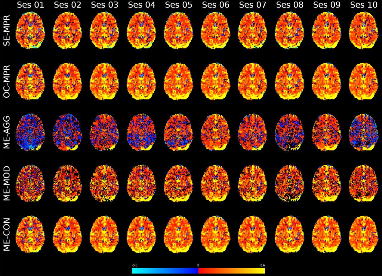
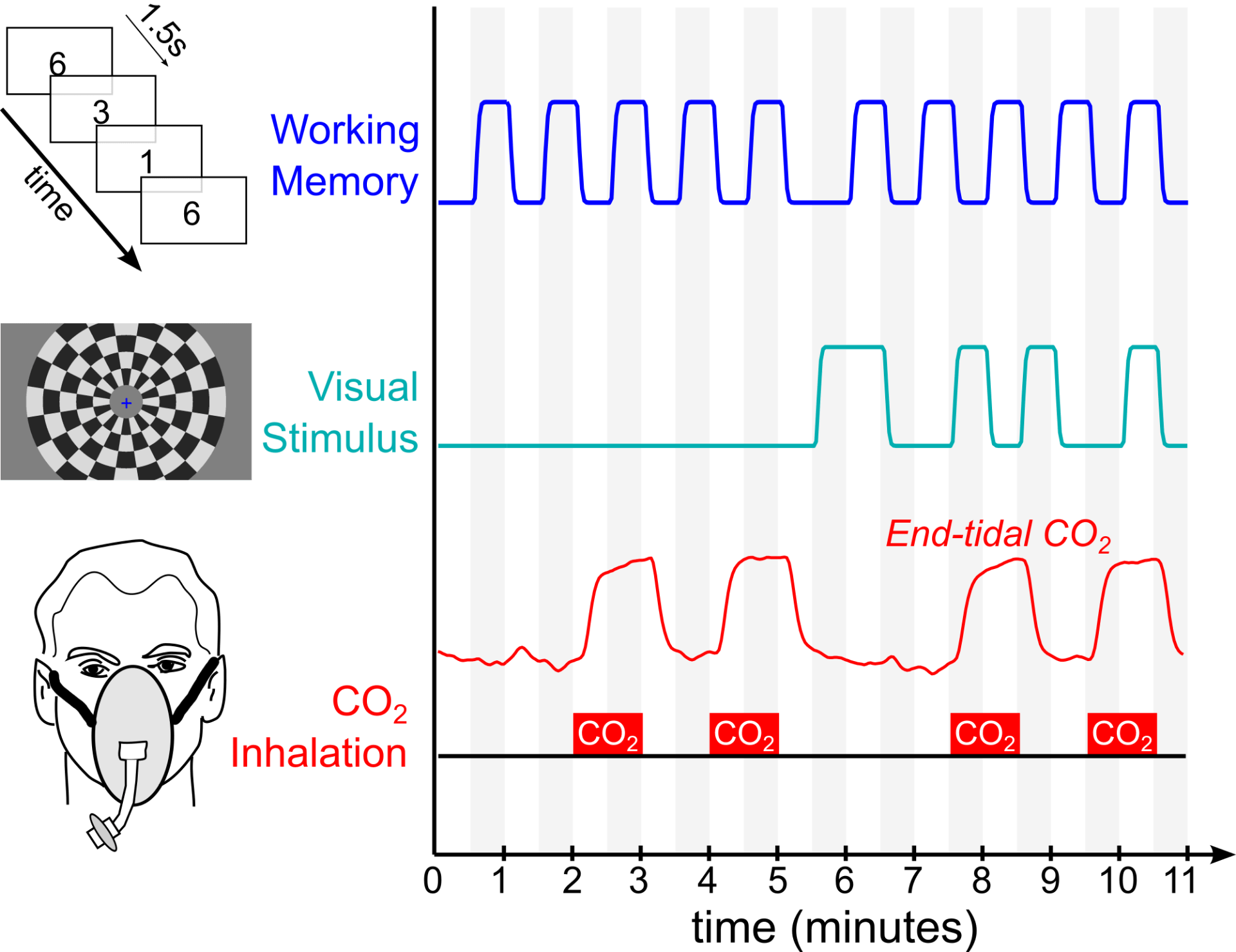
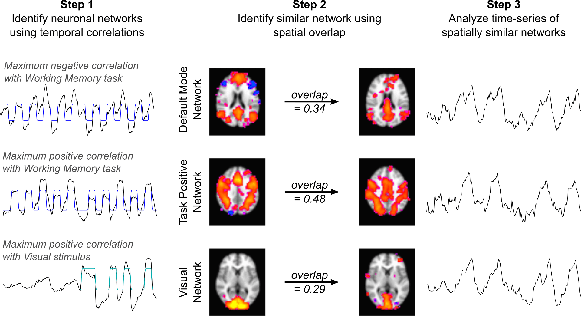
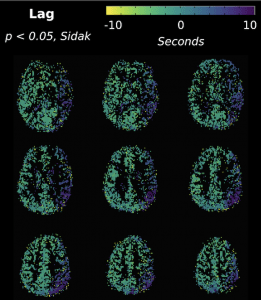
Congratulations to postdoctoral research fellow Rachael Stickland and colleagues on our publication in Neuroimage, titled A practical modification to a resting state fMRI protocol for improved characterization of cerebrovascular function.

By simply adding a few short breath-holds or deep breaths to the start of a typical resting state fMRI acquisition, we can achieve much more robust maps of cerebrovascular reactivity (CVR, the dilatory responsiveness of blood vessels) as well as the temporal delay of this response. We show that this practical approach can identify atypical cerebrovascular function, and that it is essential to understand both the amplitude and timing of the vascular response to accurately characterize pathology. This approach is easy to incorporate into traditional fMRI experiments and could help determine how vascular function and neural function are related in disease.

Find the (open access) full paper here!

New review paper on mapping CVR without gas challenges

Check out our latest publication in Frontiers Physiology, titled “Cerebrovascular Reactivity Mapping Without Gas Challenges: A Methodological Guide”. This work was lead by the excellent Joana Pinto, now a postdoc in Oxford, and covers breathing task methodology and resting state approaches for measuring CVR. This article is part of a special Frontiers “research topic” on Imaging CVR: Physiology, Physics, and Therapy.

You can find the open access article here.

Molly joins the Aperture team as Handling Editor

We are excited to announce that Aperture, the new online journal affiliated with the Organization for Human Brain Mapping, has just opened for submissions, and Molly has been invited to act as a founding Handling Editor for this exciting new format for scientific publishing. Aperture is an open-access, peer-reviewed, online journal created by members of OHBM to share and promote research beyond the traditional PDF, looking for research that will enhance, innovate, and advance neuroscience. We will publish traditional articles (research reports and reviews) but also other “research objects”, including tutorials, workshops, processing pipelines, software, simulations, computational notebooks, and datasets. For more details on the journal, submission process, and the editorial team, check out the new Aperture website.

Two new publications in collaboration with SPiN lab

Announcing the availability of two new publications in collaboration with the SPiN lab, lead by Cesar Caballero-Gaudes at the Basque Center on Cognition, Brain and Language in Spain! Both focus on the preprocessing and analysis of breath-hold fMRI data to measure cerebrovascular reactivity (CVR) and hemodynamic lag:

BCBL PhD student Stefano Moia and ANVIL postdoc Rachael Stickland co-authored an IEEE paper describing our lagged Generalized Linear Model approach for estimating the temporal delay between systemic carbon dioxide changes and the local BOLD fMRI response, mapping CVR and lag at the voxel level. The approach simultaneously fits other fMRI confounds, such as head motion, to better isolate the physiologic effects of interest. Download the pdf here!

Using the BCBL EuskalIBUR dataset, which uniquely studies breath-hold CVR across 10 repeated weekly MRI sessions, a new preprint in BioRxiv demonstrates the benefits of multi-echo data and compares multi-echo ICA denoising strategies for reliably mapping CVR and lag. Download the pdf here!

CVR maps for an example subject, scanned weekly for 10 weeks. Each row reflects a different use of multi-echo fMRI data, either combining information across the echo times or using the relationship across echo times and Independent Component Analysis (ICA) to differentiate the CVR effects from highly collinear noise confounds.

Research featured at ISMRM Annual Meeting

Recent work from the lab, lead by first author Dr. Rachael Stickland, was featured in the closing highlights session of the annual meeting of the International Society for Magnetic Resonance in Medicine. The meeting, held virtually for the first time, brought together over 5 thousand clinicians and scientists in the field of MRI and medical imaging for a week of talks, poster presentations, and virtual discussion. Dr. Stickland’s work was selected and discussed by Dr. Kevin Murphy (Cardiff University Brain Research Imaging Center), and demonstrates our lab’s proposed technique for characterizing the responsiveness of brain blood vessels using a simple, feasible, breathing task protocol during scanning. Congrats to all the authors!

Congratulations to Apoorva Ayyagari on completing her Masters Degree!

The Bright Lab celebrates Apoorva Ayyagari’s completion of her MS degree in Biomedical Engineering at Northwestern! After 2 excellent years as a core member of our lab, Apoorva successfully defended her MS thesis, titled “Understanding noise in spinal cord BOLD fMRI data with a breath-hold paradigm to investigate feasibility of studying vascular reactivity“. In this work, she rigorously assesses cardiac and respiratory noise, motion confounds, and co-linearity between different physiologic signals in spinal cord imaging data. Spinal cord fMRI data are notoriously challenging to work with due to all of these artifactual signals, and this thesis reflects excellent progress in understanding and modeling these factors. Apoorva’s many contributions in the lab will continue to benefit our ongoing projects in mapping cerebrovascular reactivity amplitude and latency, standardizing how physiologic data is collected during MRI scanning, preprocessing spinal cord fMRI data, and quantifying relationships between different sources of signal variance in these data. Although we are extremely sad to see her go, we wish Apoorva the very best as she starts her new job in human factors engineering in Evanston. Congrats Apoorva!

Demonstrating network-level interactions between neural and vascular function

Our most recent work demonstrating that the brain’s vasculature is regulated to mimic neural networks is now published in Neuroimage: https://doi.org/10.1016/j.neuroimage.2020.116907. This work was performed in collaboration with Prof. Kevin Murphy’s lab at Cardiff University Brain Research Imaging Center in the UK.

Using fMRI, we simultaneously administer two neural paradigms (a working memory task and a visual stimulus) and one “vascular” paradigm that dilates blood vessels systemically (inhaled CO2). We then averaged together 30 fMRI datasets and used Independent Component Analysis to decompose the average dataset into network components. We readily identify three “neural networks” that show strong temporal correlation with the neural stimulus paradigms. These represent the Default Mode Network, Task Positive Network, and Visual Network – three robust and commonly observed functional brain networks expected to be activated or deactivated by our neural paradigms. However, we also see three additional components with similar network structure, and these three networks predominantly reflect the vascular stimulus design.

Our results demonstrate, for the first time, pairs of spatially similar neural and vascular brain networks. This suggests that the brain’s vasculature may be regulated to support specific brain networks, which must be taken into account to interpret fMRI studies of functional connectivity.

Dr. Rachael Stickland receives NUCATS pilot grant

Congratulations to postdoctoral fellow Dr. Rachael Stickland on her success in the NUCATS Pilot Grant program! Over the coming year, Dr. Stickland will be assessing a multi-parametric imaging protocol focused on cerebrovascular function, testing reliability in healthy participants then translating into patients. This project will use the RespirActâ„¢ to deliver carbon dioxide in a carefully controlled, breath-by-breath basis during concurrent MRI scanning to characterize the associated vasodilatory response. More information about NUCATS can be found on their website.