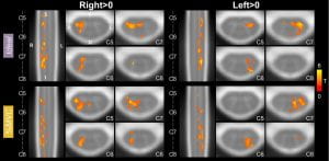
 Hand-grasping is an important and clinically significant daily function, but more work is needed to understand the associated spinal cord neural activity. In this study, participants performed a hand-grasping task with both hands and spinal cord fMRI motor activation was modeled with two methods: (1) a typical idealized block design (Ideal) and (2) based on recorded grasp force normalized to individual maximum voluntary contraction (%MVC). Robust detection of hand-grasping activity was shown in the spinal cord, highly lateralized ipsilateral to the side of the task. In addition, the impact of sample size, number of fMRI runs, and spatial smoothing on activation estimates was explored. Overall, we emphasize the importance of a task that is well-controlled within and across participants. Using individually calibrated tasks in people with motor impairments will be especially beneficial to compare across groups.If you want to learn more, please check out the paper here!
Papers
Exploring variability in spinal cord fMRI processing
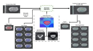 How reliable are our current techniques for processing spinal cord fMRI data? In a recently published paper, Dr. Mark Hoggarth and Max Wang explore how person-to-person variability in the contouring of fMRI spinal cord masks affects downstream analyses. Using masks drawn by 8 raters of varying experience to inform co-registration to standard template space, they identify spatial differences in the accuracy of registration related to underlying image contrast and rater experience. At the group-level, spatial differences were present in activation maps although no systematic effect on overall activation level was identified. This work characterizes how variability in manually contoured masks propagates to results in spinal cord fMRI, demonstrating that standardization of processing pipelines and improving image acquisition should be prioritized.
This work was performed in collaboration with Dr. Kenneth Weber at the Stanford Systems Neuroscience and Pain Lab.
You can find the full access paper here!
New paper on the correlation between baseline blood flow and vascular reactivity in the brain
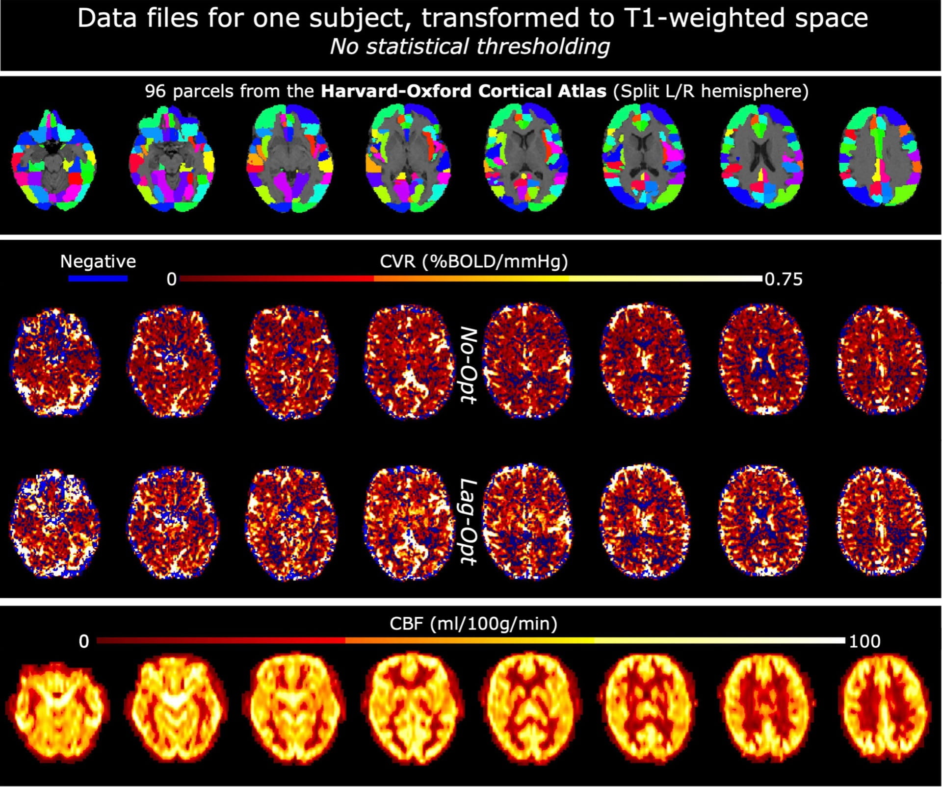 A recent paper from the lab, led by Dr. Rachael Stickland, identifies a positive correlation between baseline cerebral blood flow (CBF) and Blood Oxygenation Level Dependent cerebrovascular reactivity (BOLD-CVR), measured with MRI. This positive correlation is seen between and within subjects and is strengthened by the addition of a breathing task to a resting-state acquisition and by optimizing for hemodynamic lag effects in CVR modeling. This work demonstrates how two analytical factors affect the observed relationship
 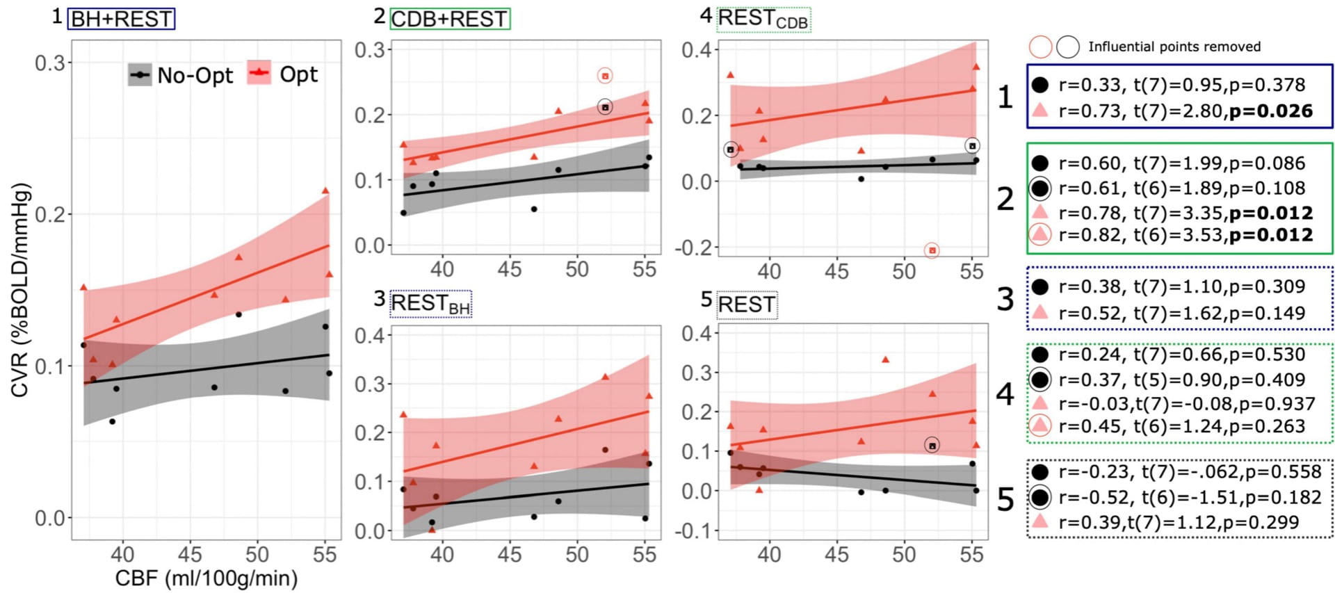 between baseline CBF and BOLD-CVR in healthy populations; the relationship becomes even more relevant to understand when interpreting results in
populations with altered vascular baselines or impaired cerebrovascular reserve.
This work was performed in collaboration with Dr. César Caballero-Gaudes’ lab at the Basque Center on Cognition, Brain and Language in Spain.
Check out the paper here!
Conference paper on visualization tool for assessment of spinal cord fMRI data quality
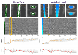 Check out our conference paper that was presented at the IEEE Engineering in Medicine and Biology Conference and published online in December 2021! This paper details our method, developed by PhD candidate Kimberly Hemmerling, for visualization of complex spinal cord fMRI data as heatmaps. This work was inspired by a visualization technique designed for brain fMRI data (Power et al., 2017). Our heatmaps can be visualized alongside motion or physiological traces that may be used to identify coincidence of signal variation. The technique may be easily integrated into a preprocessing pipeline to visualize the effect of a preprocessing step, identify spatially or temporally varying artifacts, or to assess general data quality.Check out the work here, and our GitHub repository (BrightLab-ANVIL/spinalcordplot), which contains the code and example data necessary to generate the plots.
New review paper on mapping CVR without gas challenges
Check out our latest publication in Frontiers Physiology, titled “Cerebrovascular Reactivity Mapping Without Gas Challenges: A Methodological Guide”. This work was lead by the excellent Joana Pinto, now a postdoc in Oxford, and covers breathing task methodology and resting state approaches for measuring CVR. This article is part of a special Frontiers “research topic” on Imaging CVR: Physiology, Physics, and Therapy.
Two new publications in collaboration with SPiN lab
Announcing the availability of two new publications in collaboration with the SPiN lab, lead by Cesar Caballero-Gaudes at the Basque Center on Cognition, Brain and Language in Spain! Both focus on the preprocessing and analysis of breath-hold fMRI data to measure cerebrovascular reactivity (CVR) and hemodynamic lag:
BCBL PhD student Stefano Moia and ANVIL postdoc Rachael Stickland co-authored an IEEE paper describing our lagged Generalized Linear Model approach for estimating the temporal delay between systemic carbon dioxide changes and the local BOLD fMRI response, mapping CVR and lag at the voxel level. The approach simultaneously fits other fMRI confounds, such as head motion, to better isolate the physiologic effects of interest. Download the pdf here!
Using the BCBL EuskalIBUR dataset, which uniquely studies breath-hold CVR across 10 repeated weekly MRI sessions, a new preprint in BioRxiv demonstrates the benefits of multi-echo data and compares multi-echo ICA denoising strategies for reliably mapping CVR and lag. Download the pdf here!
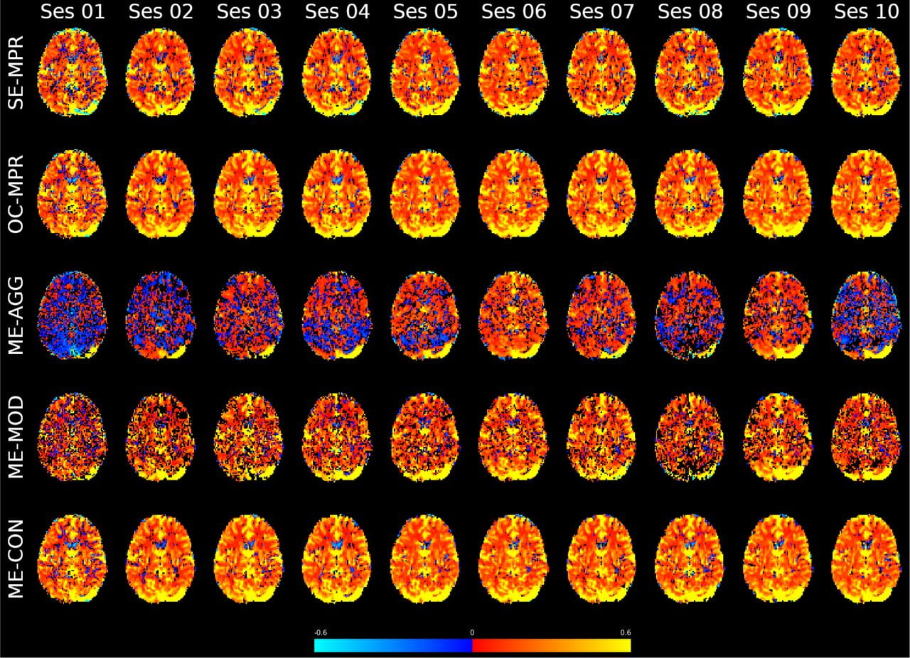 CVR maps for an example subject, scanned weekly for 10 weeks. Each row reflects a different use of multi-echo fMRI data, either combining information across the echo times or using the relationship across echo times and Independent Component Analysis (ICA) to differentiate the CVR effects from highly collinear noise confounds.
Demonstrating network-level interactions between neural and vascular function
 Our most recent work demonstrating that the brain’s vasculature is regulated to mimic neural networks is now published in Neuroimage: https://doi.org/10.1016/j.neuroimage.2020.116907. This work was performed in collaboration with Prof. Kevin Murphy’s lab at Cardiff University Brain Research Imaging Center in the UK.
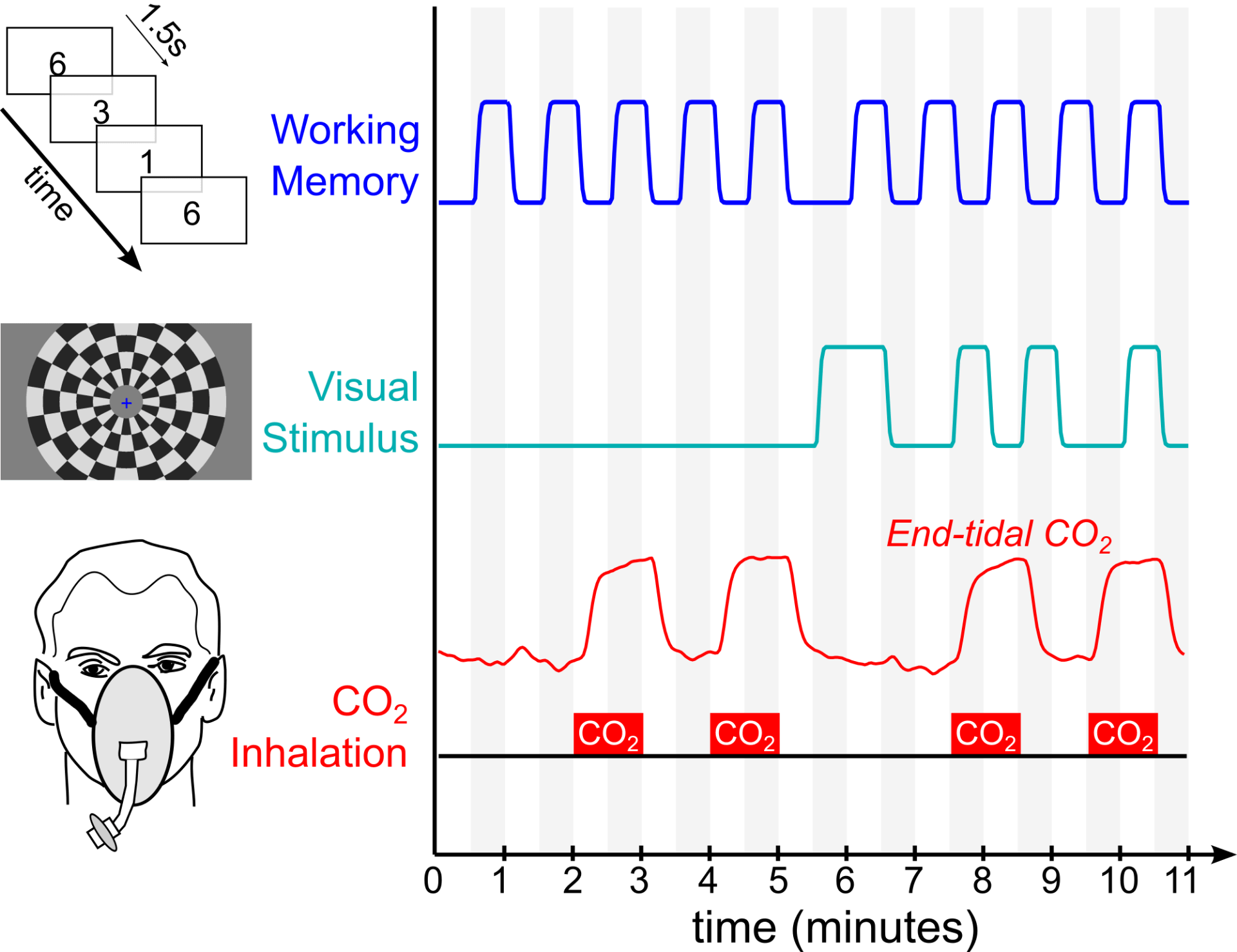
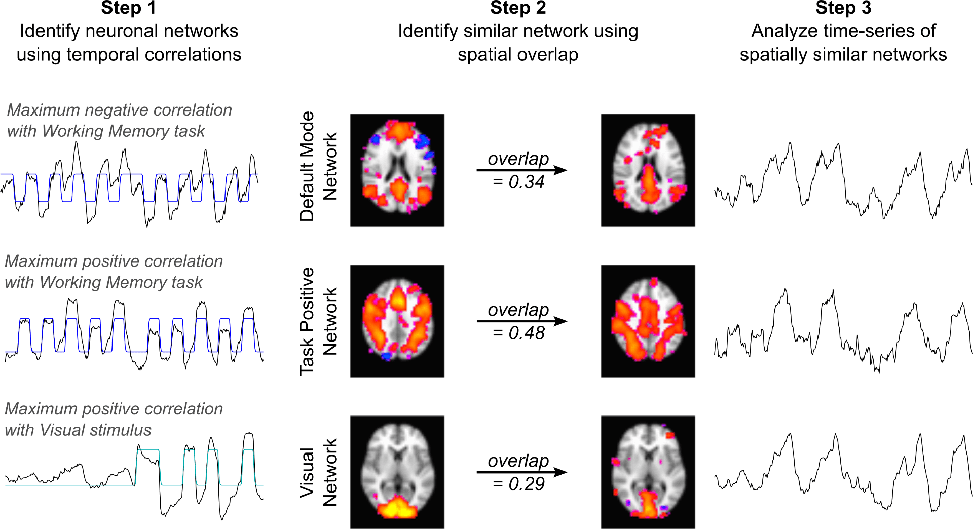
Using fMRI, we simultaneously administer two neural paradigms (a working memory task and a visual stimulus) and one “vascular” paradigm that dilates blood vessels systemically (inhaled CO2). We then averaged together 30 fMRI datasets and used Independent Component Analysis to decompose the average dataset into network components. We readily identify three “neural networks” that show strong temporal correlation with the neural stimulus paradigms. These represent the Default Mode Network, Task Positive Network, and Visual Network – three robust and commonly observed functional brain networks expected to be activated or deactivated by our neural paradigms. However, we also see three additional components with similar network structure, and these three networks predominantly reflect the vascular stimulus design.
Our results demonstrate, for the first time, pairs of spatially similar neural and vascular brain networks. This suggests that the brain’s vasculature may be regulated to support specific brain networks, which must be taken into account to interpret fMRI studies of functional connectivity.
Ultra-high field MRI gives new insight into perfusion of lesions in Multiple Sclerosis
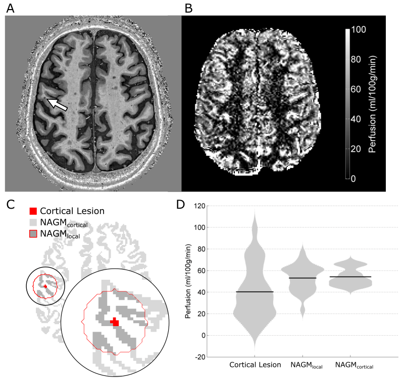 In our latest paper in European Radiology, we use ultra-high field MRI to explore the clinical usefulness of an emerging technique for measuring perfusion that doesn’t involve the injection of contrast agents. Instead, in Arterial Spin Labeling (ASL) scans, we magnetically label the blood and then image it as it flows into the tissue, allowing us to quantify perfusion. We developed this technique for a specific clinical application: to better understand the role of small cortical lesions in Multiple Sclerosis (MS).
In this technical development report, we present an optimized ultra-high-field ASL MRI acquisition that achieves high spatial resolution and low signal distortion. We assess the feasibility of using this imaging strategy to measure perfusion in MS cortical lesions, and demonstrate that our approach is sensitive to focal hypoperfusion in these small lesions. Because ASL MRI is safe and non-invasive, this type of imaging approach may facilitate the longitudinal study of acute lesion formation and development with frequent repeated scanning, allow us to test new therapeutic strategies, and give us better understanding of the heterogenous disease course in MS.
This excellent work was primarily accomplished by Rich Dury and Yasser Falah as part of their PhD research. For more details, the manuscript is available (open access) here: https://rdcu.be/8n4G
New paper on multiparametric physiological imaging
 A new article from the Bright Lab, titled “Multiparametric measurement of cerebral physiology using calibrated fMRI“, has been accepted for publication in NeuroImage.
In the context of a calibrated fMRI experiment, we simultaneously characterize BOLD, blood flow, blood volume, tissue oxygen extraction, oxidative metabolism, and vascular reactivity throughout the brain. Rather than focus on a single aspect of brain physiology, considering the relationships of multiple physiological parameters allows more accurate and precise characterization of neurovascular health in a range of clinical applications. This paper is part of a special issue on Physiological MRI, in collaboration with Paula Croal, Nic Blockley, and Dan Bulte at the University of Oxford.