Ultra-high field MRI gives new insight into perfusion of lesions in Multiple Sclerosis

In our latest paper in European Radiology, we use ultra-high field MRI to explore the clinical usefulness of an emerging technique for measuring perfusion that doesn’t involve the injection of contrast agents. Instead, in Arterial Spin Labeling (ASL) scans, we magnetically label the blood and then image it as it flows into the tissue, allowing us to quantify perfusion. We developed this technique for a specific clinical application: to better understand the role of small cortical lesions in Multiple Sclerosis (MS).

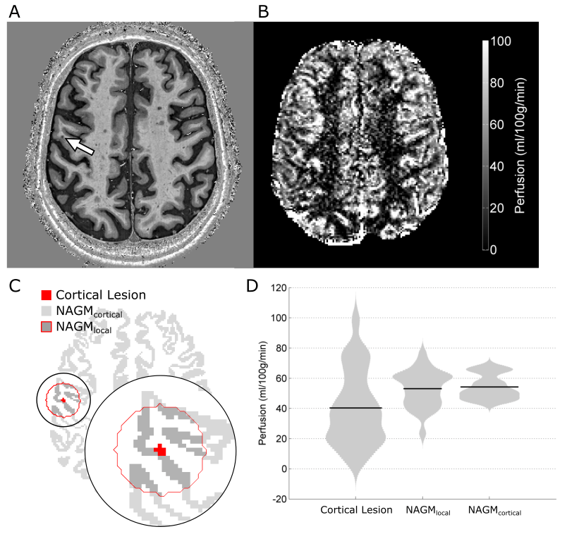
In this technical development report, we present an optimized ultra-high-field ASL MRI acquisition that achieves high spatial resolution and low signal distortion. We assess the feasibility of using this imaging strategy to measure perfusion in MS cortical lesions, and demonstrate that our approach is sensitive to focal hypoperfusion in these small lesions. Because ASL MRI is safe and non-invasive, this type of imaging approach may facilitate the longitudinal study of acute lesion formation and development with frequent repeated scanning, allow us to test new therapeutic strategies, and give us better understanding of the heterogenous disease course in MS.

This excellent work was primarily accomplished by Rich Dury and Yasser Falah as part of their PhD research. For more details, the manuscript is available (open access) here: https://rdcu.be/8n4G

Career Development Award for PI Molly Bright

We are excited to announce that Molly Bright has received an Interdisciplinary Rehabilitation Engineering Research Career Development (K12) Award! The IREK12 program recruits and trains scholars with engineering and other quantitative backgrounds to become successful rehabilitation scientists in basic, translational and/or clinical research. The award will fund research in adapting fMRI methodology for use in pediatric brain injury, constructing and validating new models of the BOLD signal in individual children, and then applying these techniques to study atypical motor systems in pediatric-onset hemiplegia. For more information about the IREK12 program, see the consortium’s webpage.

Welcome Kristina Zvolanek to the lab!

ANVIL welcomes Kristina Zvolanek, a new PhD student in Biomedical Engineering. Kristina joins us from the University of Nebraska at Lincoln, where she earned her B.S. in Biological Systems Engineering and already accomplished a first-author paper in Medical Physics. As an undergraduate, she worked with Dr. Greg Bashford in the Biomedical Imaging and Biosignal Analysis lab, where she used Transcranial Doppler ultrasonography to measure cerebral blood flow patterns in children. At Northwestern, she will continue to pursue her interests in pediatric imaging and rehabilitation, adapting and applying functional and physiological MRI techniques to study neurovascular properties of the brain in pediatric-onset hemiplegia. She’s already familiar with Chicago and Northwestern, having previously done a summer research project with Dr. Sabrina Lee in the Neuromuscular Biomechanics Lab, and has gotten a jump-start on her graduate research in our lab prior to the start of classes in September. We are extremely excited to have Kristina on the team, and look forward to working with her in this exciting area of pediatric and rehabilitative neuroimaging!

Open position for a Postdoctoral Fellow

Update: An offer has been extended and accepted. Please see the Openings page for details on current positions.

We are excited to announce an opening for a full-time Postdoctoral Fellow to develop advanced MRI techniques to assess the interaction of neural activity and vascular physiology. These techniques will be applied in several collaborative themes, studying novel therapeutics in rehabilitation, exploring brain-body relationships in aging, and characterizing motor impairment and plasticity in children and adults following stroke. There is opportunity and support for the Fellow to initiate and lead new directions in basic and clinical research. This position is for a minimum of 2 years with the possibility to extend.

For further details, please see the full description on the Openings page. Interested applicants are encouraged to get in touch with Prof. Bright prior to submitting an application. It may also be possible to arrange for an informal discussion in person during the ISMRM annual meeting in Paris. (Applications received prior to July 2, 2018 will receive full consideration.)

We look forward to hearing from you!

New paper on multiparametric physiological imaging

A new article from the Bright Lab, titled “Multiparametric measurement of cerebral physiology using calibrated fMRI, has been accepted for publication in NeuroImage.

In the context of a calibrated fMRI experiment, we simultaneously characterize BOLD, blood flow, blood volume, tissue oxygen extraction, oxidative metabolism, and vascular reactivity throughout the brain.  Rather than focus on a single aspect of brain physiology, considering the relationships of multiple physiological parameters allows more accurate and precise characterization of neurovascular health in a range of clinical applications. This paper is part of a special issue on Physiological MRI, in collaboration with Paula Croal, Nic Blockley, and Dan Bulte at the University of Oxford.

View the full paper here.

Bright Lab opens at Northwestern University!

The Bright Lab of Applied Neurovascular Imaging is moving to Northwestern University on January 1st, 2018. Based downtown in the Chicago Campus, the lab is embedded in the applied clinical research facilities of Physical Therapy and Human Movement Sciences, right on the Magnificent Mile, and links with the dedicated neuroimaging scanners at the Centre for Translational Imaging. Several openings for PhD students and post-doctoral research fellows will be announced in the new year. Stay tuned for updates as we apply advanced MRI techniques for studying cerebral physiology in a range of exciting new clinical directions!